Paper 2 / Signed biological evidence
Signed Dopaminergic Asymmetry Tracks Contralateral Motor Laterality in Parkinson's Disease
The amount of asymmetry does not tell us which side is affected. Retaining its direction distinguishes opposite biological states and exposes disagreement between imaging and examination. It helps describe the present more clearly than it predicts the future.
Actual PPMI records / Four different information states
Which side, according to which measurement?
Cases A and B show opposite imaging-motor directions. Case C has nearly symmetric imaging despite an asymmetric examination. In Case D the two sources disagree. These are measured records, including cases where added imaging did not help.
The complete records remain available in the four patient dossiers.
Sign convention, case selection and model inputs
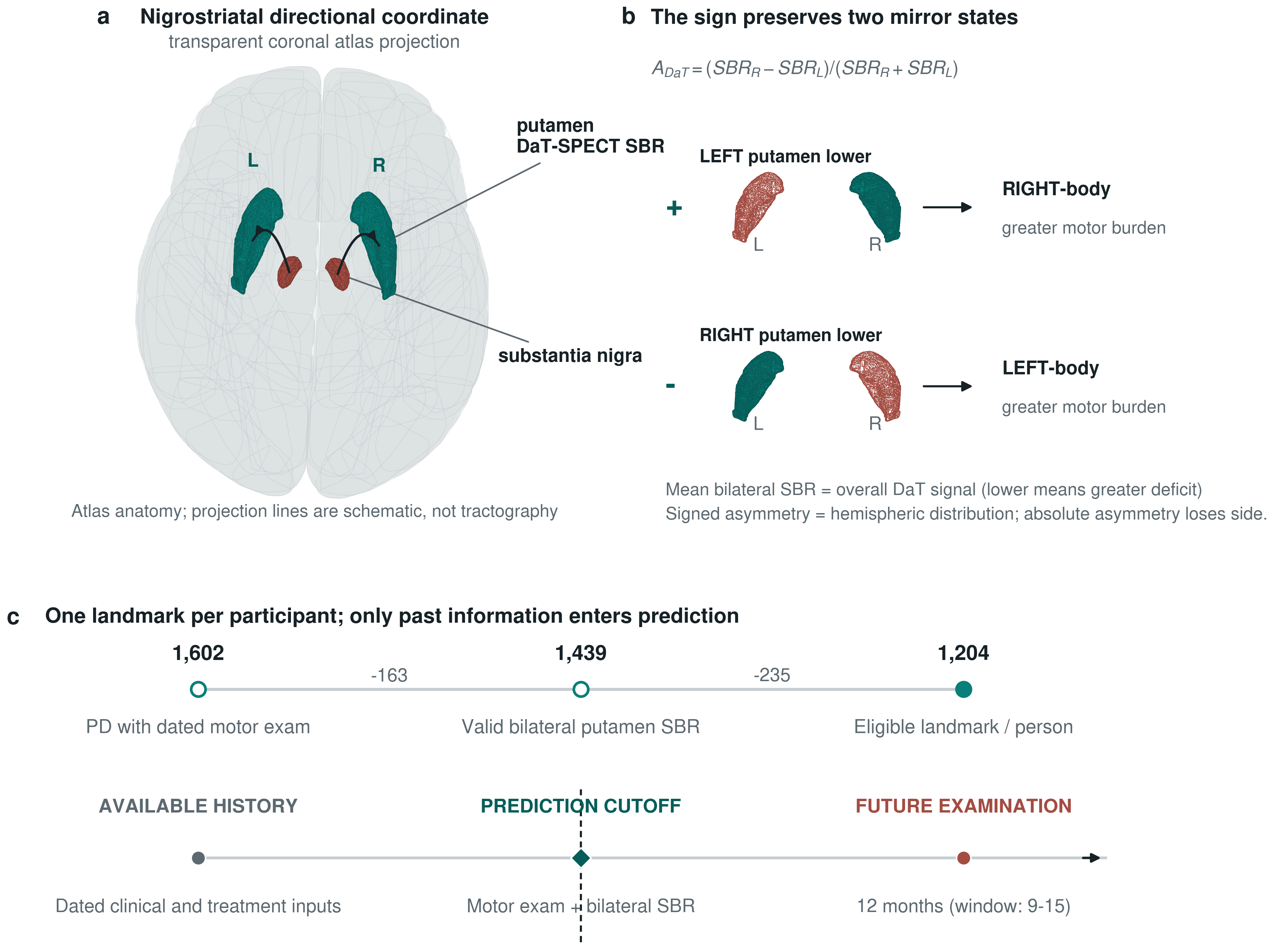
Signed imaging is (right SBR - left SBR) / (right SBR + left SBR). Signed motor laterality is (right paired motor sum - left paired motor sum) / (right paired motor sum + left paired motor sum). Thus positive imaging means relatively lower left putamen binding; positive motor laterality means greater right-body burden. Lower binding on one side is not a measurement of global disease severity.
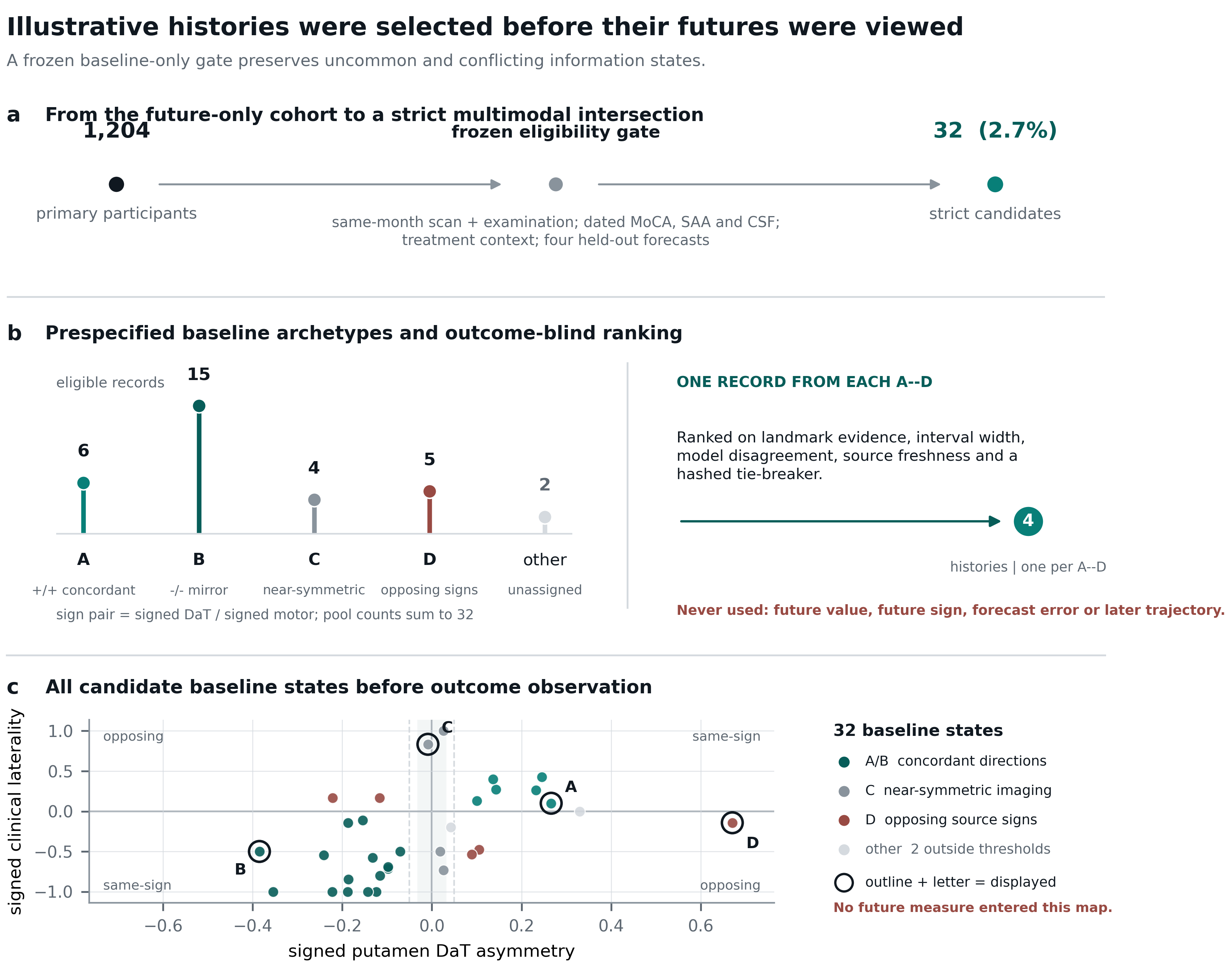
The four frozen cases came from the 32-record strict multimodal pool within 1,204 longitudinal participants. Baseline state, source freshness, held-out model disagreement, interval width and a hashed tie-breaker were used, not the individual's future value or forecast error. A and B are not severity-matched. The fuller selection map follows below.
Clinical scores, demographics, examination state and LEDD enter the clinical model; imaging is added in the comparison models. MoCA, SAA and CSF values are contextual annotations, not inputs to these focused forecasts. These sources were also examined in separate exploratory context comparisons; those tests did not establish a molecular explanation for the directional differences. CSF percentiles are relative to the 32-record illustration pool, not clinical reference ranges.
| Measurement | Scale and meaning | Role here |
|---|---|---|
| Putamen SBR | Regional imaging: dopamine-transporter specific-binding ratio | Mean, magnitude and signed forecast comparisons |
| Paired motor scores | Clinical examination: matched left/right MDS-UPDRS III items | Current state and separately dated future endpoint |
| LEDD and examination state | Treatment record: levodopa-equivalent dose and ON/OFF documentation | Clinical-model inputs, with missingness retained |
| MoCA | Clinical cognition: screening score out of 30 | Display and exploratory context, not a forecast input |
| SAA, CSF alpha-synuclein and NfL | Fluid biomarkers: seeding assay, protein measurements and neurofilament light | Display and exploratory context, not causal explanations |
The 90% interval and its median belong to a separate conformalized quantile-regression model. They must not be attached to the four ridge-model point forecasts. Points on the history trace are actual examinations; connecting segments are not measured intervening states or a simulation of disease.
Directional coordinate
A side-aware representation retains anatomical meaning
Positive imaging values are fixed to relatively lower left putamen binding; positive clinical values are fixed to greater right-body burden. The orientation is defined before analysis and is carried unchanged across PPMI, S4, and future outcomes.
Outcome-blind selection audit
The illustrative histories were chosen before their futures were viewed
Thirty-two of 1,204 future-only participants met the strict same-month and multimodal completeness gate. Four baseline information states were defined in advance. One record was selected from each state using only landmark evidence, source freshness, model disagreement, interval width, and a hashed tie-breaker.
Patient-state evidence explorer
Read each record as a sequence, not a pile of modalities
Each dossier keeps source age, bilateral anatomy, clinical side balance, observed history and four held-out model forecasts in one view. The separate quantile-model median and interval are not uncertainty bounds for the ridge forecasts. The records were selected to expose different information states, not four favorable predictions.
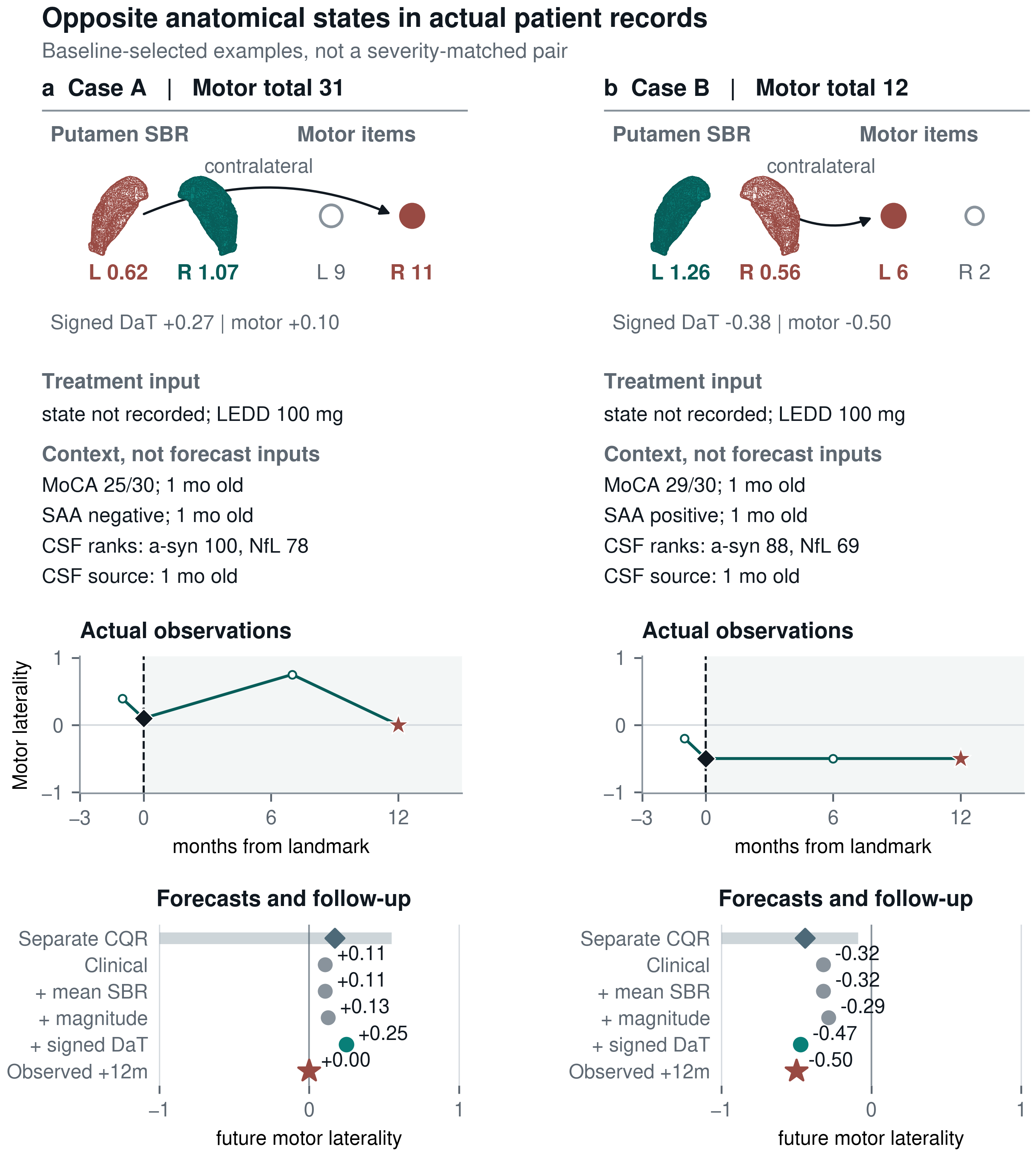
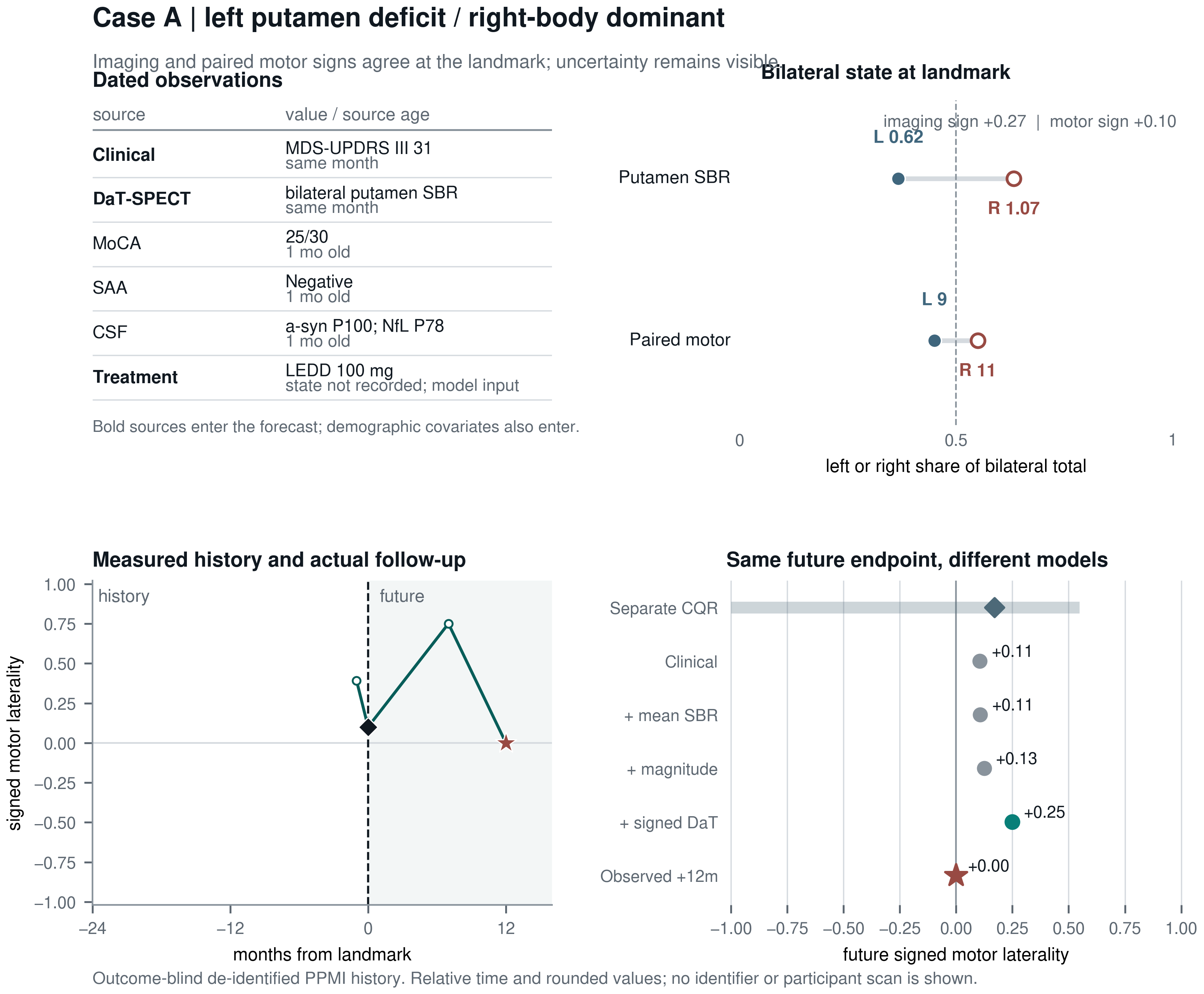
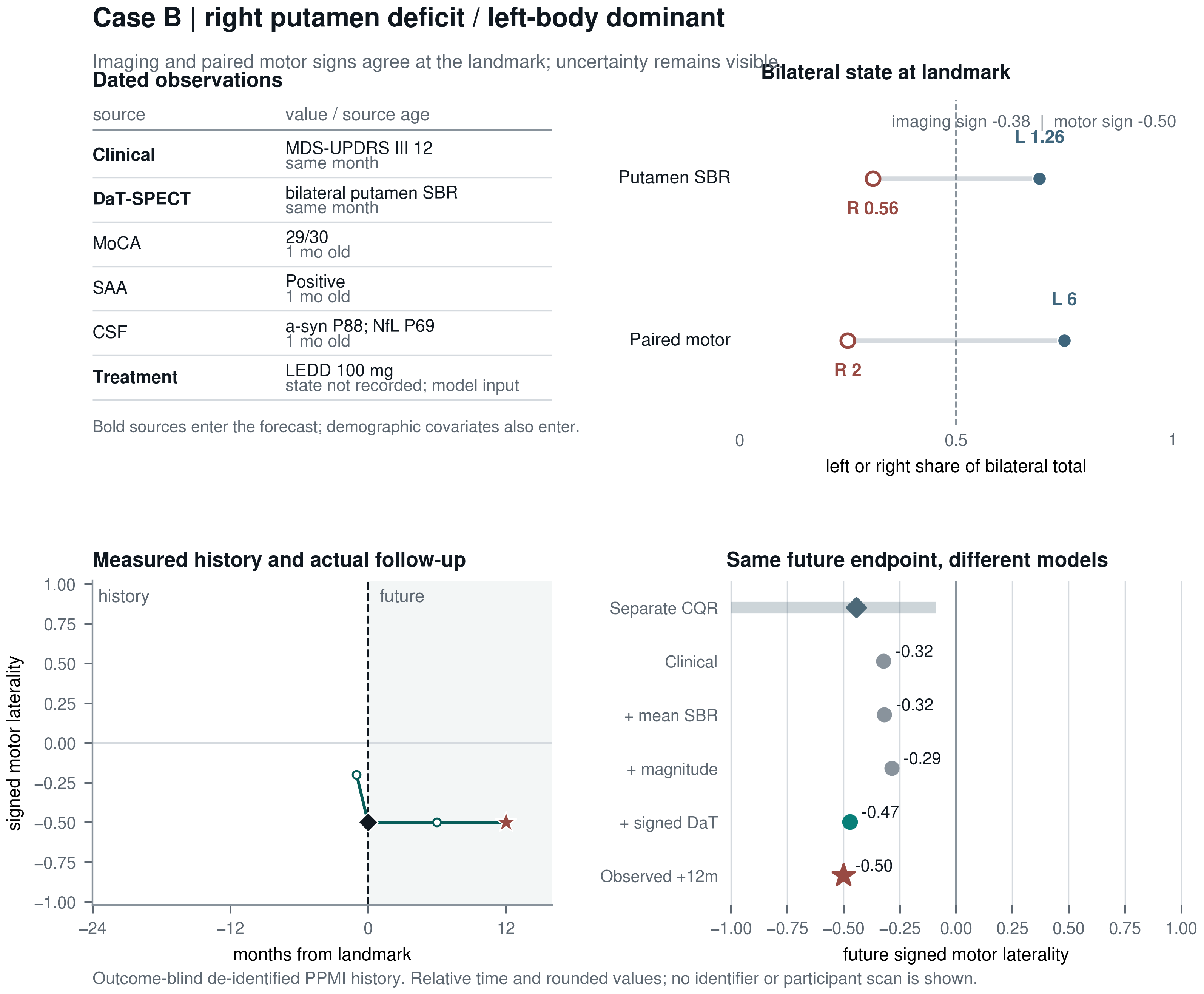
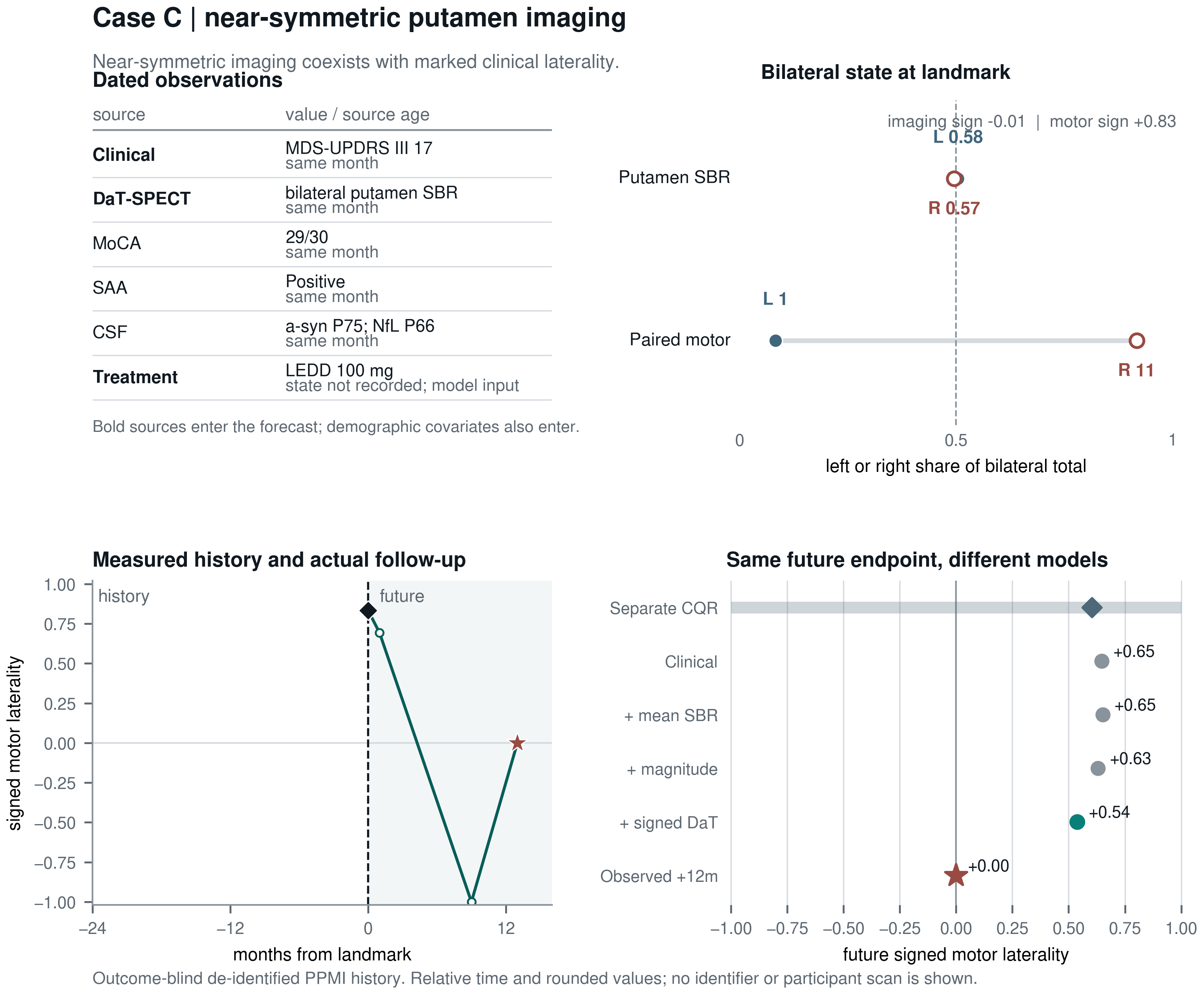
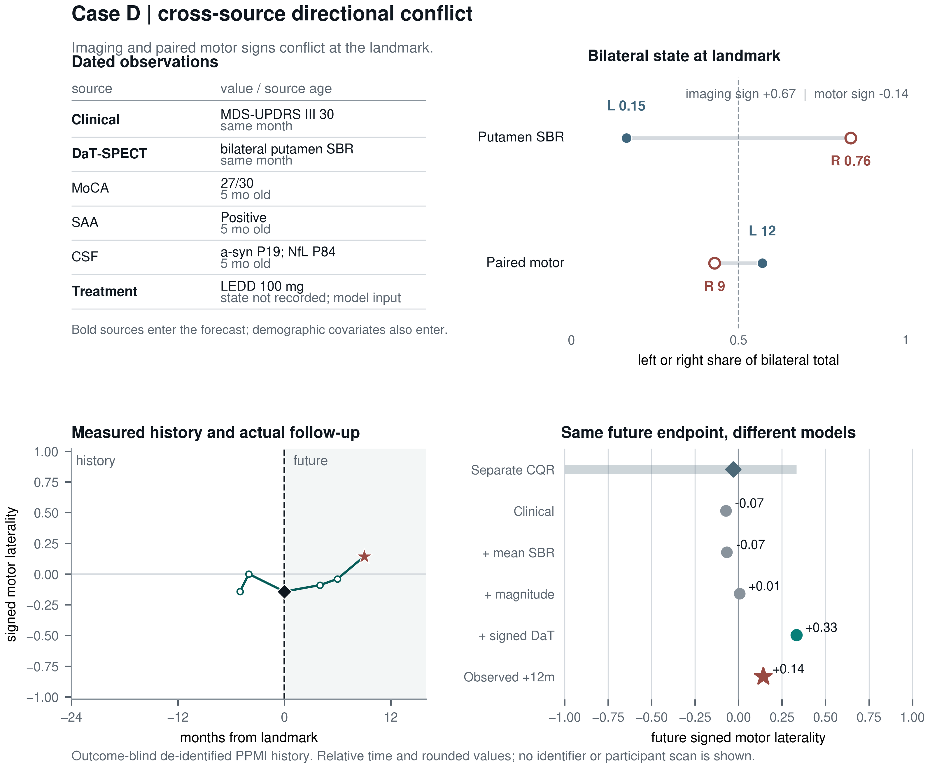
Labels are arbitrary and one-use, time is relative to each landmark, and displayed values are rounded. The dossiers contain no identifier, calendar date, source record, or participant scan. They explain information states; cohort-level analyses carry the inference.
Biological replication
Signed anatomy reproduces across independent cohorts
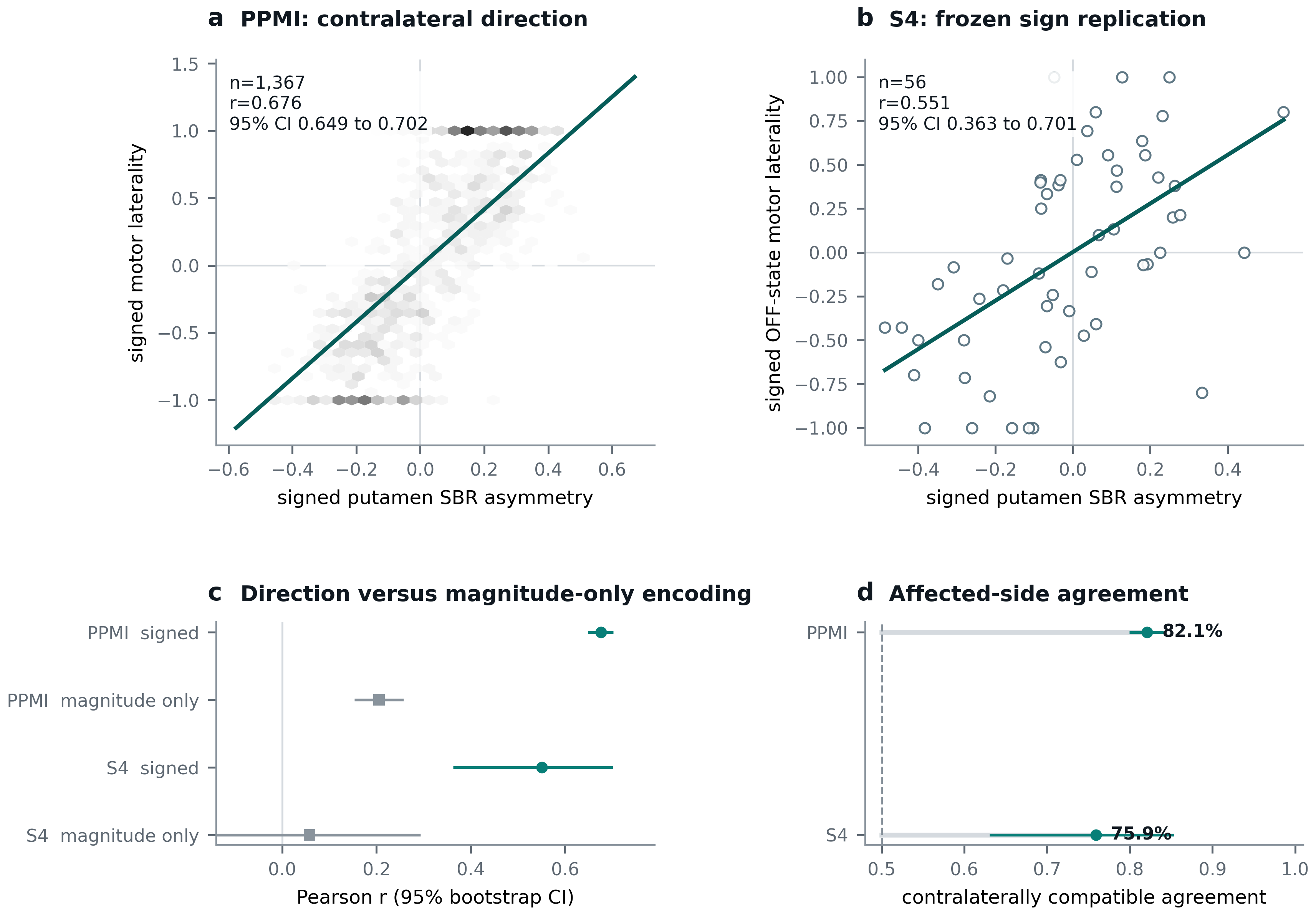
Replication supports a transferable anatomical coordinate. It does not establish a causal mechanism, clinical diagnosis, or treatment target by itself.
Future side-balance
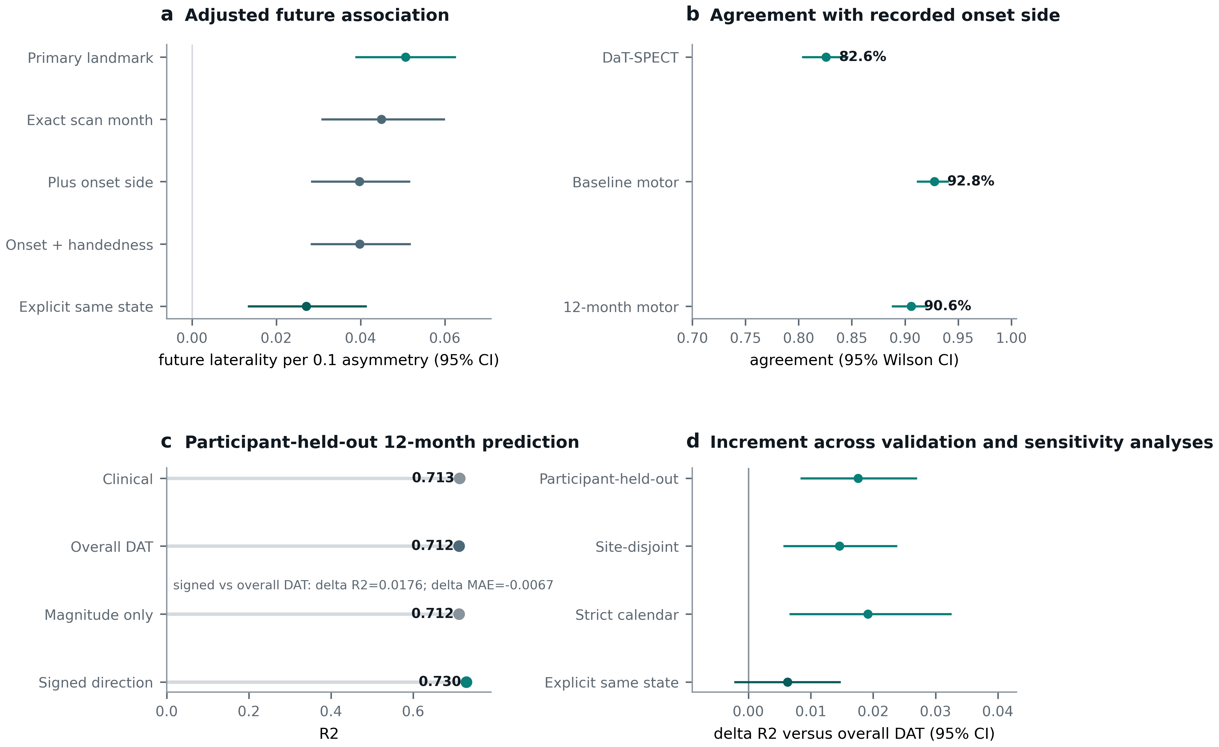
Direction carries modest longitudinal information
Making disagreement visible
Imaging and clinical direction do not always agree
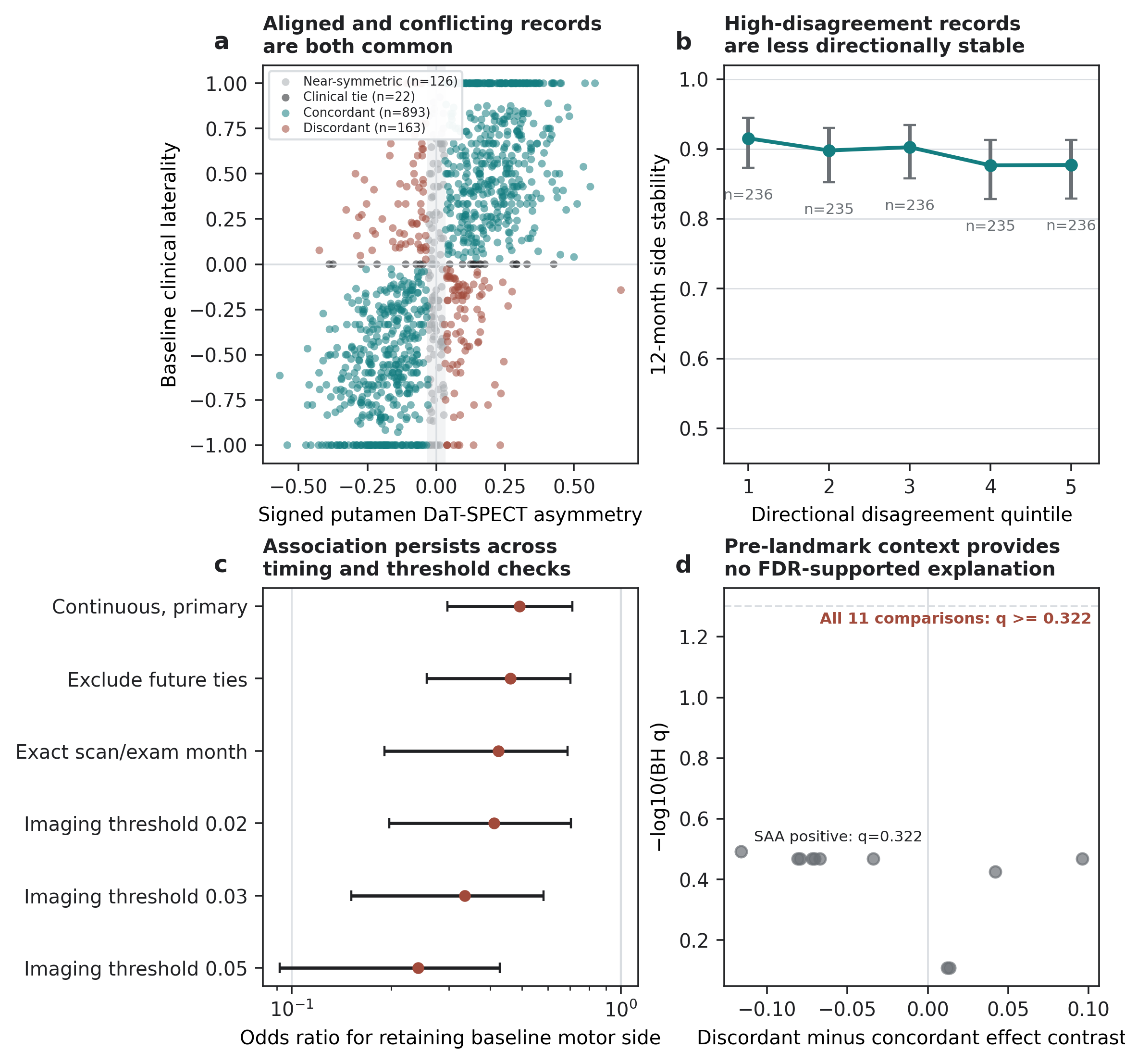
A useful state must preserve disagreement rather than forcing every source into one label. The analysis therefore separates records whose imaging and clinical signs agree, disagree, or contain weak directional evidence.
Claim boundary
What sign adds, and what it does not
Which patients would an unsigned summary merge? A secondary audit identified 365 non-overlapping patient pairs with similar total motor scores, mean putamen binding and absolute asymmetry, but opposite imaging signs. In 281 pairs, the clinical signs were opposite too. Retaining direction keeps these biological states distinguishable.
These are not 365 misdiagnoses. The clinical reference already includes clinical laterality. In the full future-outcome comparison, binary side accuracy was 90.2% for the clinical model and 89.8% after adding signed imaging: no improvement. Nor did the earlier matched-profile analysis establish that secondary biomarkers explain future divergence. More informative representation, better prediction and biological explanation remain separate questions.
| Question | Result | Interpretation |
|---|---|---|
| Contralateral biological anatomy | Replicated | Signed putamen asymmetry tracks opposite-side motor burden in PPMI and S4 |
| Future continuous side-balance | Limited information | Association persists, but predictive improvement is small |
| Binary side classification | No improvement | Sign does not improve every outcome representation |
| Axial motor prediction | No improvement | The coordinate is anatomically specific rather than a global severity score |
| Clinical utility or treatment benefit | Not tested | Prospective response and treatment-policy validation remain necessary |
Contribution
- A frozen signed DaT-SPECT coordinate
- Independent cross-cohort biological replication
- Future-only participant-held-out laterality testing
- Outcome-blind patient histories that retain disagreement and uncertainty
Not claimed
- Global Parkinson disease progression prediction
- A causal or calibrated mechanism probability
- Treatment response or treatment recommendation
- Deployment-ready clinical utility
Connected research / Different questions, different claims
From patient phenotypes toward mechanism-aware evidence
- 01 / Pathway context
CVC pathway-anchored clustering
Organizes structural, microstructural and dopaminergic imaging by predefined biological pathways.
- 02 / Patient phenotypes
Posterior-aware phenotyping
Addresses clinical heterogeneity and uncertainty in patient representations.
bioRxiv preprint - 03 / Biological specificity
Signed dopaminergic laterality
Retains direction, tests contralateral agreement and preserves unresolved conflict.
- 04 / Validity limits
Longitudinal validity gates
Tests when a patient-state forecast is informative and when its clinical claim must stop.
Our earlier integrated genetic, molecular and wearable-biomarker study (medRxiv preprint) provides complementary multimodal stratification context. It is distinct from CVC's pathway-anchored imaging study.
This is a research progression, not a single jointly validated diagnostic pipeline. Patient-specific intervention simulation and treatment selection remain future work. The AI4PD program provides the broader systems context; these pages replay fixed research examples, not live clinical predictions.
Data: PPMI; access to underlying participant data follows the source study's application process. Code is available from the authors on request; the paper-specific repository is pd-signed-laterality (access required). Putamen outlines adapt the NIH 3D Human Reference Atlas "Brain, Male" (3DPX-020960), based on the Allen Human Reference Atlas under CC BY 4.0, rendered here as neutral two-dimensional views. They are not patient scans.